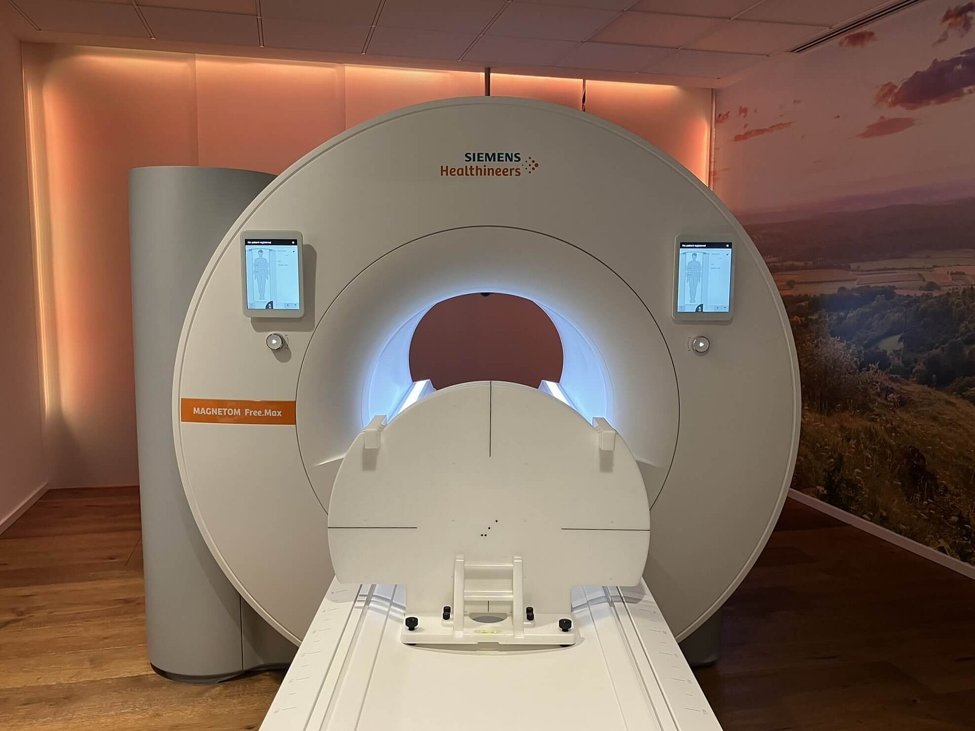
Detect distortion of MR images
The THETIS 3D MR Distortion Phantom serves to detect possible distortion of MR images reliably and quickly. It contains small markers arranged in a square grid that provide a strong, localized MR signal. By determining the amount of distortion of the square grid or signal loss on the MR image, possible image distortion can be detected by the user. The phantom is aligned to the isocenter or the image center of the MR unit using MR laser systems from LAP and can be aligned easily and quickly using the integrated leveling aids.
MR in radiation therapy
There are different reasons for distortions in MR imaging. The most important ones are distortions due to gradient non-linearities, B0 inhomo-geneities and susceptibility artefacts caused by the imaged patient or phantom. All MRI manufacturers
provide methods and algorithms for distortion correction to minimise these effects.
In clinical practice, residual image distortion needs to be below acceptable limits. A large field phantom such as THETIS is able to detect residual distortions from gradient non-linearities or B0 main magnet inhomogeneties. Its embedded silicon markers arranged in a grid help to visualise the limited geometry away from the magnet isocentre and prevent a potential inaccurate view of organs at the outer area of MRI image.
Having a tool on hand that conveniently detects distortions contributes to making clinical life safer and easier. The result leads to better patient treatment, which is the goal in RT.
Find the protocols for THETIS MR Distortion Phantom test on the SIEMENS Healthineers website.
Technical data
| Weight (total) | 15,5 kg |
|---|---|
| Dimensions phantom plate | 10 mm length, 666 mm width, 486 mm height |
| Weight phantom plates | 3,6 kg |
| Dimensions levelling plate | 400 mm length, 350 mm width, 20 mm height |
| Weight levelling plate | 2,4 kg |

